• Home
  • Portfolio
  • Non-Extraction Correction of a Severe Anterior Open Bite with Closure of Large Posterior Spaces

Non-Extraction Correction of a Severe Anterior Open Bite with Closure of Large Posterior Spaces

Anterior open bite is widely regarded as one of the most difficult malocclusions to correct and one of the most likely to relapse. The decisive question, however, is not mechanical but diagnostic: is the open bite skeletal or dental in origin? A skeletal open bite driven by a divergent growth pattern behaves very differently from a dental open bite in which the jaw bases are essentially normal and the incisors have simply been displaced. Treating the second as if it were the first leads to unnecessary surgery or unnecessary extractions; treating the first as if it were the second leads to relapse.

This patient, a 24-year-old woman, presented with a severe anterior open bite that extended posteriorly into the premolar region, producing a characteristic Y-shaped separation of the lateral occlusal planes. Her ABO Discrepancy Index was 37, of which the anterior open bite alone accounted for 26 points. The situation was complicated by a retained root of tooth 16, which had allowed a large space to develop in the maxillary right posterior segment, and by the retention of all four third molars — 18 and 38 in particular held the occlusal fulcrum far posteriorly, which actively sustained the open bite.

Despite a clinical picture in which most conventional indicators pointed toward extraction, the case was resolved without extracting a single premolar. Three coordinated strategies made this possible: coordinating the upper and lower arch forms, systematically relocating the occlusal fulcrum forward, and using infrazygomatic crest and mandibular buccal shelf miniscrews to control the vertical dimension during retraction and distalization. Active treatment took 32 months and finished with an ABO Cast-Radiograph Evaluation score of 15, comfortably within the passing threshold of 34 for a case of this complexity.

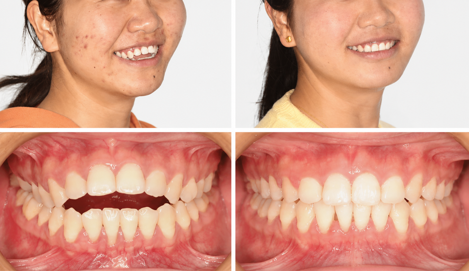
Figure 1. Pre-treatment and post-treatment comparison of facial appearance and anterior occlusion
 
 
Patient Information
 
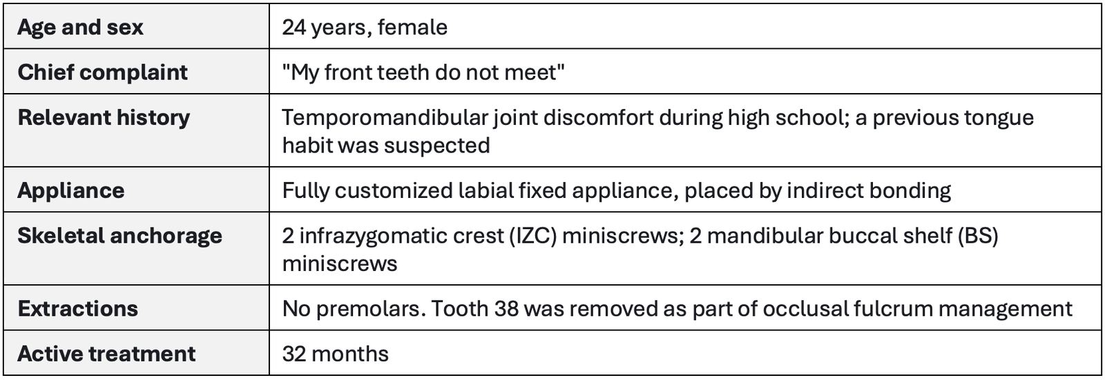
Table 1. Basic background information
 
Clinical Examination

Extraoral Findings

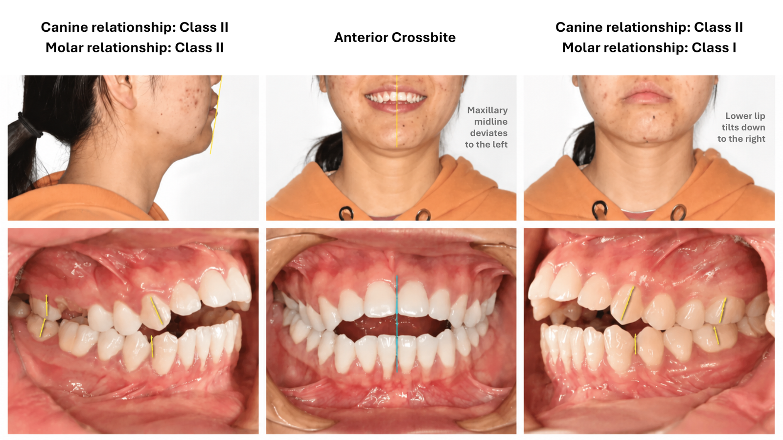
  • Downturned oral commissures at rest.
  • Uneven commissure height on smiling, producing an asymmetric smile line.
  • The commissure recess (buccal corridor) on the right side was larger than on the left.

Intraoral Findings

  • Anterior open bite involving the incisor region and extending into the premolar region.
  • Right side: canine relationship distal; molar relationship distal.
  • Left side: canine relationship distal; molar relationship neutral (Class I).
  • Upper dental midline deviated to the left.
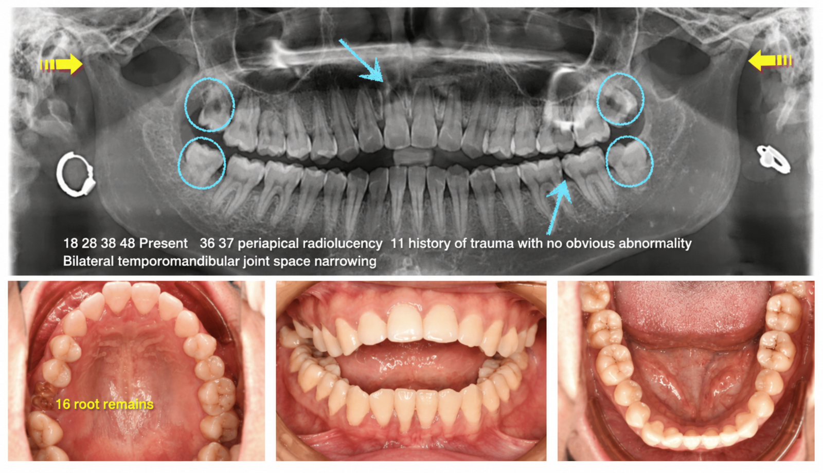
  • Tooth 16 present as a retained root, with a large residual space in the maxillary right posterior segment.

Radiographic Findings

  • Teeth 18, 28, 38 and 48 all present.
  • Interproximal caries affecting teeth 36 and 37.
  • Tooth 11 with a history of trauma; no obvious periapical abnormality at presentation.
  • Bilateral condyles short and small in form.
  • On the lateral cephalogram, the lateral occlusal planes diverged to form a Y-shaped open bite.
Figure 2. Pre-treatment extraoral and intraoral photographic series, showing the anterior open bite, the canine and molar relationships on each side, and the leftward deviation of the upper midline
 
 
Figure 3. Pre-treatment panoramic radiograph and occlusal views

*Annotations record the four retained third molars, interproximal caries on 36 and 37, the traumatised tooth 11, the short bilateral condyles, and the retained root of 16
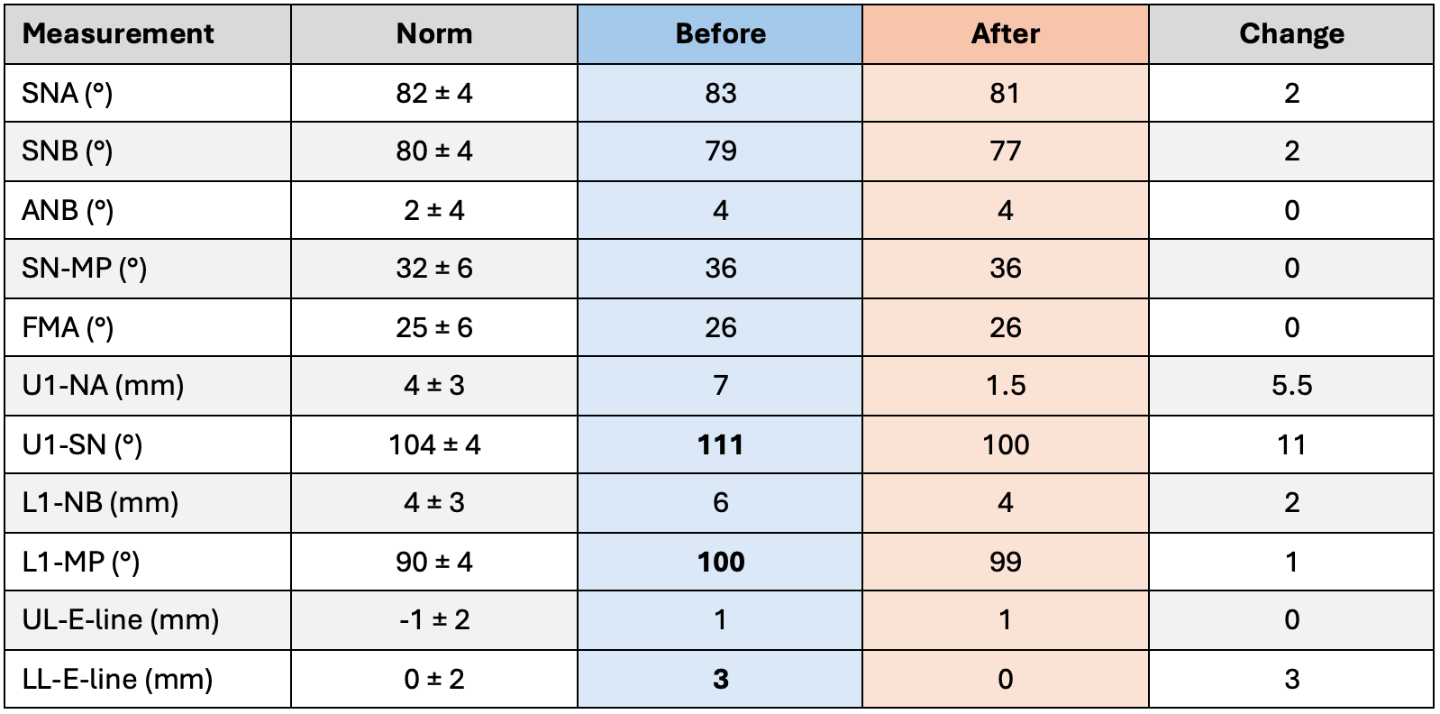
 

Table 2. Cephalometric measurements before and after treatment.

 The skeletal pattern was Class I (ANB 4°) with an average mandibular plane angle (FMA 26°, SN-MP 36°). The abnormal findings were confined to the dentition and soft tissue: both upper and lower incisors were proclined (U1-SN 111°, L1-MP 100°) and the lower lip was protrusive relative to the E-line (3 mm). This distribution — normal jaw bases with displaced incisors — identified the open bite as predominantly dental in origin.


Diagnosis

  • Angle Class I malocclusion
  • Skeletal Class I
  • Average mandibular plane angle (normodivergent)
  • Anterior open bite, dental in origin

Contributing and complicating factors:

  • Proclined maxillary and mandibular incisors.
  • Retained root of tooth 16 with a large residual posterior space.
  • Occlusal fulcrum held far posteriorly by the retained third molars 18 and 38.
  • Upper dental midline deviation to the left.
  • Interproximal caries on 36 and 37; previously traumatised tooth 11.

Case Complexity Assessment

The American Board of Orthodontics Discrepancy Index was scored before treatment.
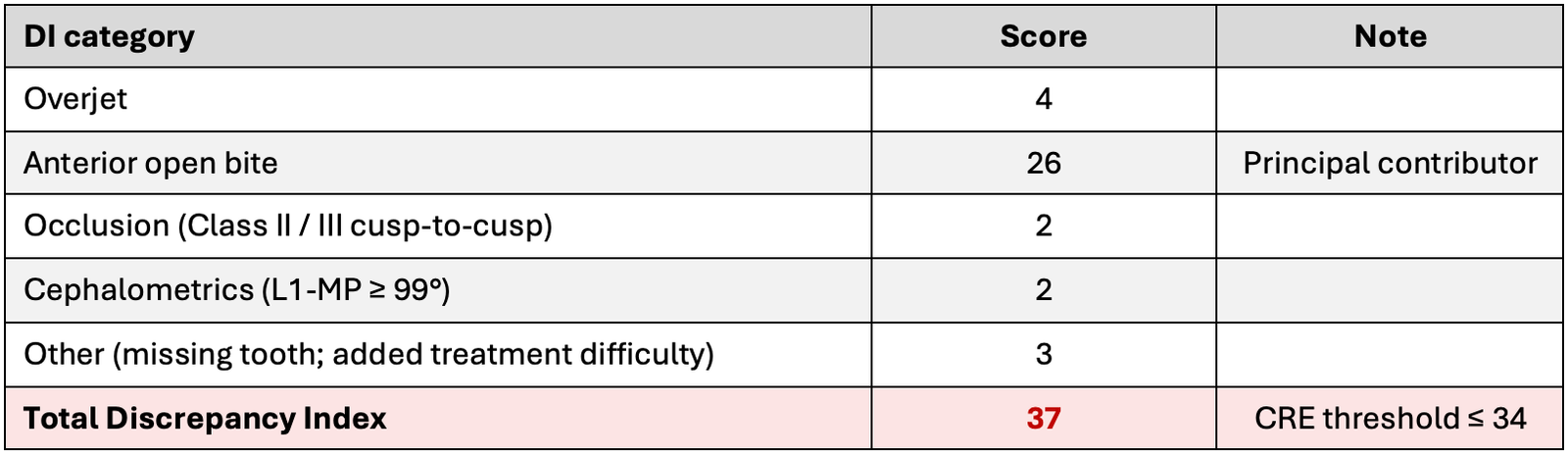
Table 3. ABO Discrepancy Index breakdown

*A DI in the 30–39 band requires a Cast-Radiograph Evaluation score of 34 or lower to pass

More than two-thirds of the total score came from the anterior open bite alone, which confirmed that the vertical problem, rather than crowding or the anteroposterior relationship, was the defining challenge of the case.


Treatment Objectives


  • Close the anterior open bite and establish a positive overbite and overjet.
  • Close the large posterior space left by the 16 retained root without prosthetic replacement.
  • Retrocline the proclined maxillary incisors and improve lip support.
  • Correct the leftward deviation of the upper dental midline.
  • Coordinate the maxillary and mandibular arch forms and eliminate the reverse curve of the lower arch.
  • Relocate the occlusal fulcrum mesially so that it no longer props the bite open.
  • Maintain the vertical dimension and avoid clockwise rotation of the mandible during retraction and distalization.
  • Establish a stable posterior occlusion and secure long-term vertical stability through appropriate retention.

Treatment Plan

Seven reference criteria were reviewed before committing to a plan.
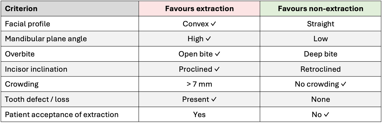
Table 4. The seven-criterion extraction decision matrix, with the patient’s status marked in each row
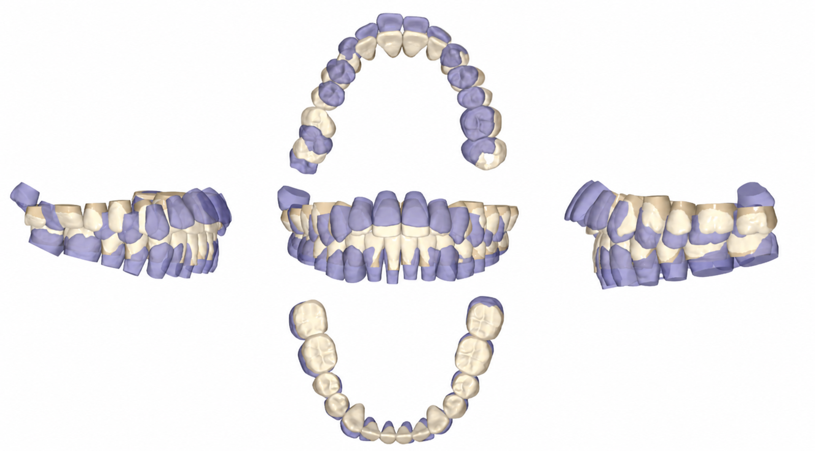
Figure 4. Digital setup simulation comparing the initial tooth positions (purple) with the planned final positions (cream), used to derive the individualized bracket prescription
 

Appliance and Bracket Design

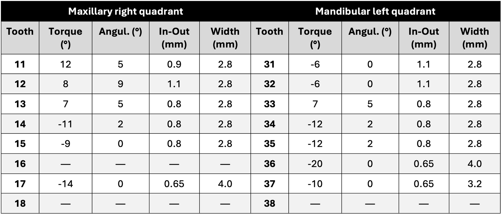
A fully customized labial bracket system was used. Every torque, angulation, in-out and bracket width value was individualized to the patient’s tooth morphology and to the target positions established in the digital setup, then transferred to the mouth with an indirect bonding tray to preserve the accuracy of that prescription. The parameters supplied in the digital bracket report are listed below.

Table 5. Individualized bracket prescription
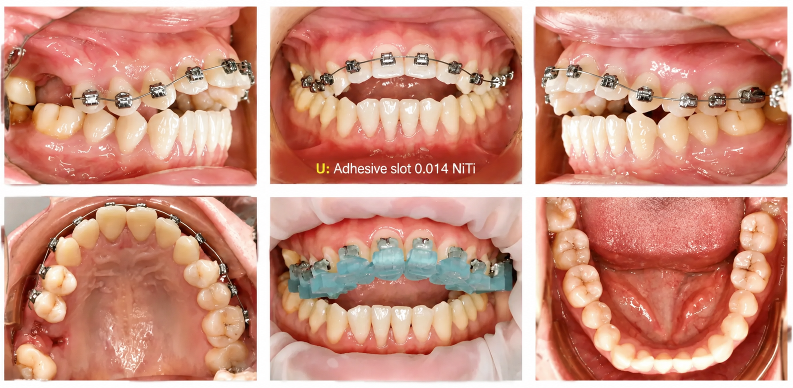
 

Figure 5. Appliance placement at 0 months

*The maxillary arch was bonded with an indirect bonding transfer tray and levelled on a 0.014 NiTi archwire

Treatment Sequence
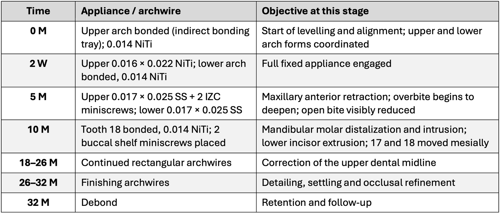
Table 6. Chronological treatment sequence over 32 months of active treatment
 
Mechanics: Three Keys to Closing the Open Bite


Key 1: Coordinate the arch forms and eliminate the reverse curve

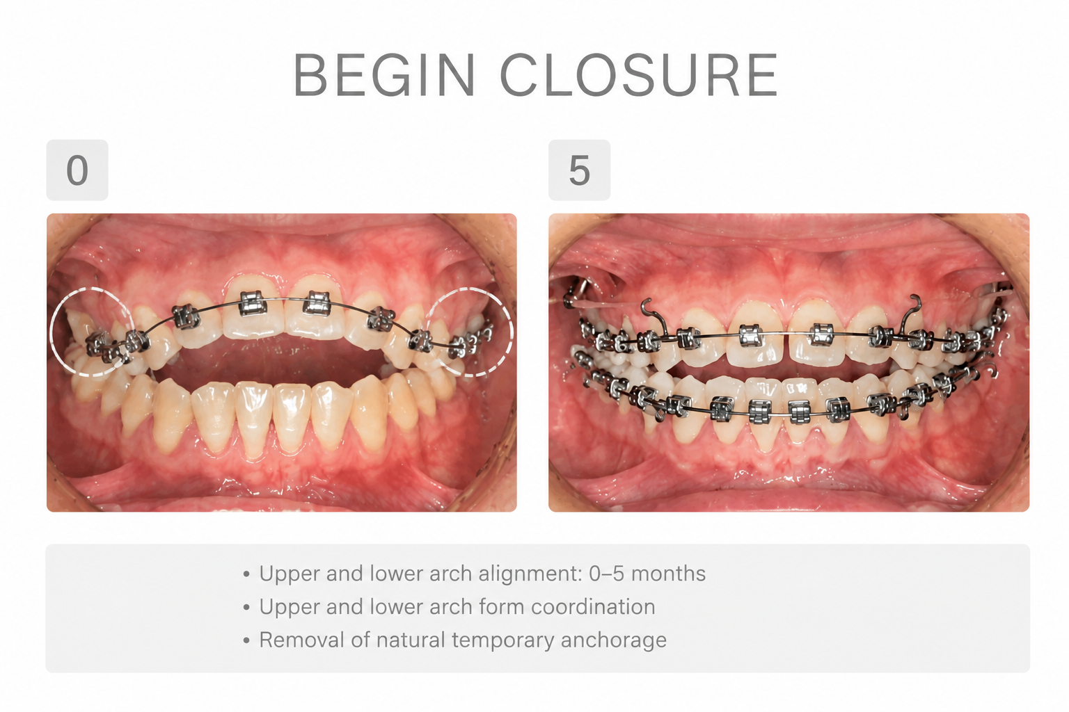
The maxillary posterior segment was narrow relative to the mandibular arch. Levelling and aligning both arches to a matched arch form during the first five months removed the natural reverse (rocking-chair) curve of the lower arch. This alone produced a visible reduction in the open bite before any skeletal anchorage was placed — which answers the question of why simple alignment improved the bite at all.

Figure 6. Arch form coordination between 0 and 5 months. Matching the upper and lower arch forms removed the reverse curve and reduced the open bite.
 

Diagnosis


Key 2: Move the occlusal fulcrum forward

With teeth 18 and 38 both present, the occlusal fulcrum sat at the very back of the arch, acting as a pivot that levered the anterior teeth apart. Tooth 38 was extracted, which left 18 without an antagonist and eliminated the distal fulcrum altogether. Teeth 17 and 18 were then moved mesially, carrying the occlusal contact point forward with them. Closing the posterior space in this direction served two purposes at once: it eliminated the residual space from the 16 retained root and moved the fulcrum to a position where it no longer propped the bite open.


Figure 7. Fulcrum management. Left: extraction of 38 leaves 18 without an antagonist and removes the distal fulcrum. Right: mesial movement of 17 and 18 carries the occlusal contact point forward.
 

Key 3: Use skeletal anchorage for vertical control

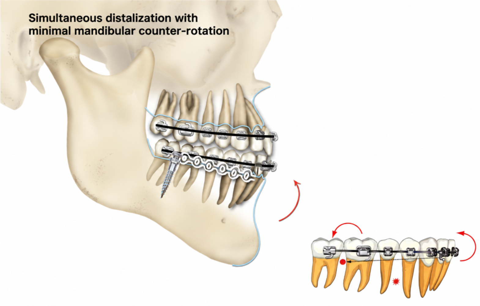
At five months, two infrazygomatic crest miniscrews were placed to retract the maxillary anterior segment. Retraction of proclined incisors deepens the overbite, and the open bite reduced accordingly. At ten months, two mandibular buccal shelf miniscrews were added to distalize and intrude the lower molars while allowing the lower incisors to extrude. En-masse distalization against buccal shelf anchorage produced a small counter-rotation of the mandibular occlusal plane, which closes an open bite instead of opening it further. The result was closure achieved with the mandibular plane angle entirely unchanged.


Figure 12. Maxillary retraction against IZC miniscrews from 5 months, with progressive deepening of the overbite at 0, 5 and 10 months.


Figure 8. Schematic of en-masse distalization against buccal shelf anchorage, producing a small counter-rotation of the mandibular occlusal plane

Treatment Outcomes


Clinical, photographic and cephalometric evaluation after 32 months of active treatment confirmed correction of every presenting concern.

Occlusion and alignment

  • The anterior open bite was completely closed, with a positive overbite and overjet established across the incisors.
  • The posterior space associated with the 16 retained root was closed by mesial movement; no prosthetic replacement was required.
  • Full arch alignment achieved with a stable, well-interdigitated posterior occlusion.
  • The upper dental midline was corrected.

Cephalometric change

  • Maxillary incisor position: U1-SN reduced from 111° to 100° (11° of uprighting) and U1-NA from 7 mm to 1.5 mm — the largest single change in the case.
  • Mandibular incisor position: L1-NB reduced from 6 mm to 4 mm; L1-MP from 100° to 99°.
  • Vertical control: SN-MP unchanged at 36° and FMA unchanged at 26°. Despite substantial retraction, distalization and molar intrusion, there was no clockwise rotation of the mandible — the single most important indicator that the open bite closure was achieved by design rather than by chance.
  • Sagittal relationship: ANB unchanged at 4°; SNA 83° to 81° and SNB 79° to 77°, preserving the Class I skeletal pattern.
  • Soft tissue: The lower lip returned to the E-line (3 mm to 0 mm), with the upper lip unchanged at 1 mm, giving a balanced lower facial profile.

ABO Cast-Radiograph Evaluation

The finished result scored 15 on the ABO Cast-Radiograph Evaluation. Recorded deductions included alignment and rotation (3), buccolingual inclination (2), occlusal contacts (4) and interproximal contacts (2). For a case with a Discrepancy Index of 37, the passing threshold is 34 — the result therefore passed with a substantial margin.

Planned restorative follow-up

  • Tooth 11: root canal therapy planned.
  • Teeth 36 and 37: composite resin restorations planned.
Figure 9. Intraoral photographs at debonding, 32 months. The open bite is closed with positive overbite and overjet, and the posterior spaces are fully closed.
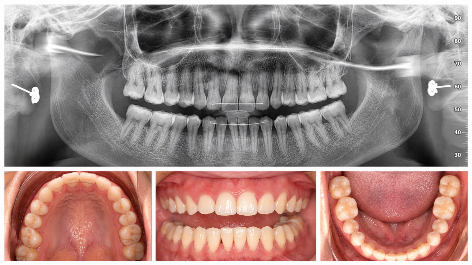
 

Figure 10. Post-treatment panoramic radiograph and occlusal views showing root parallelism and closed posterior spaces
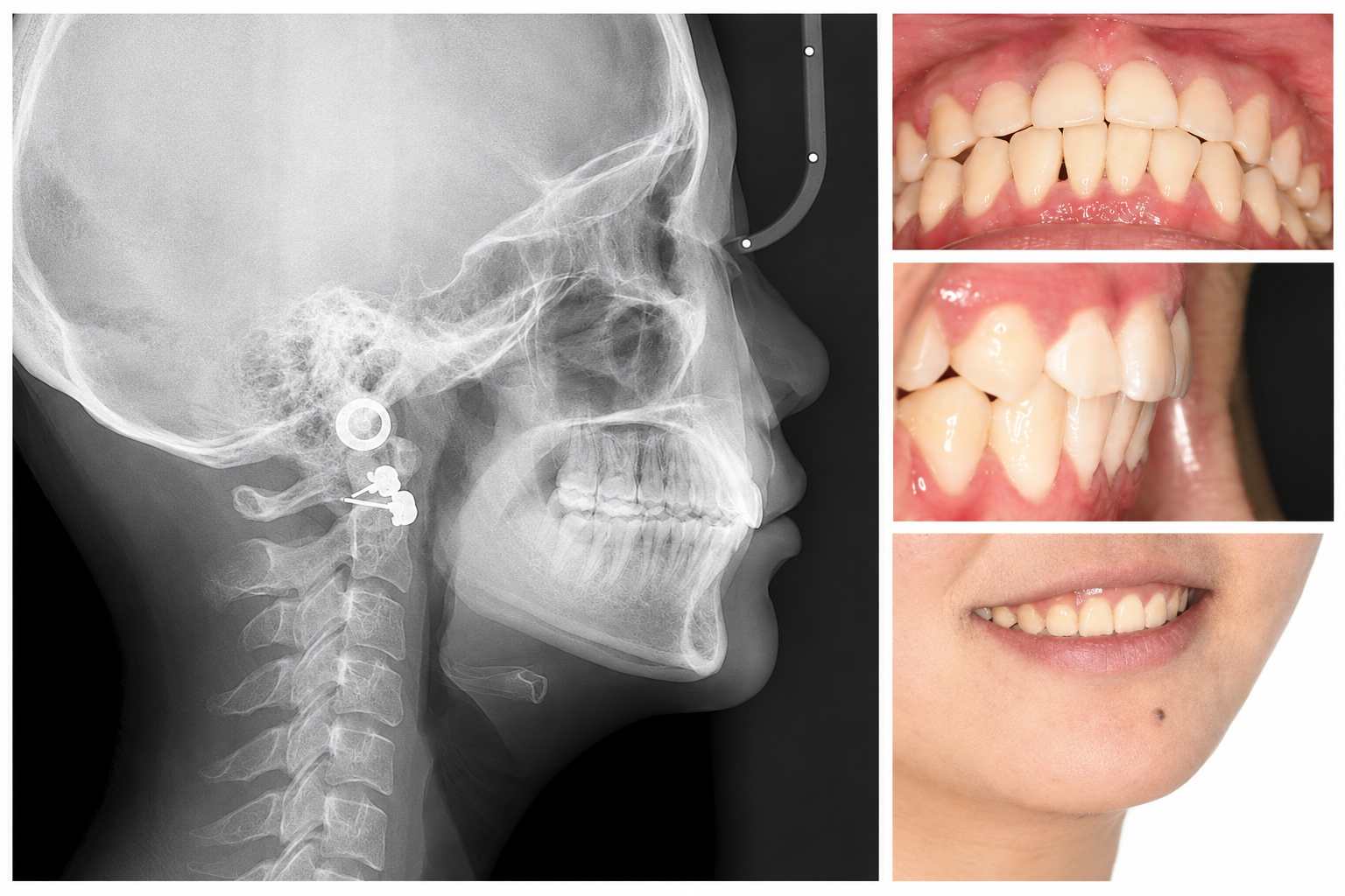
Figure 11. Post-treatment lateral cephalogram with the corresponding intraoral and smile views
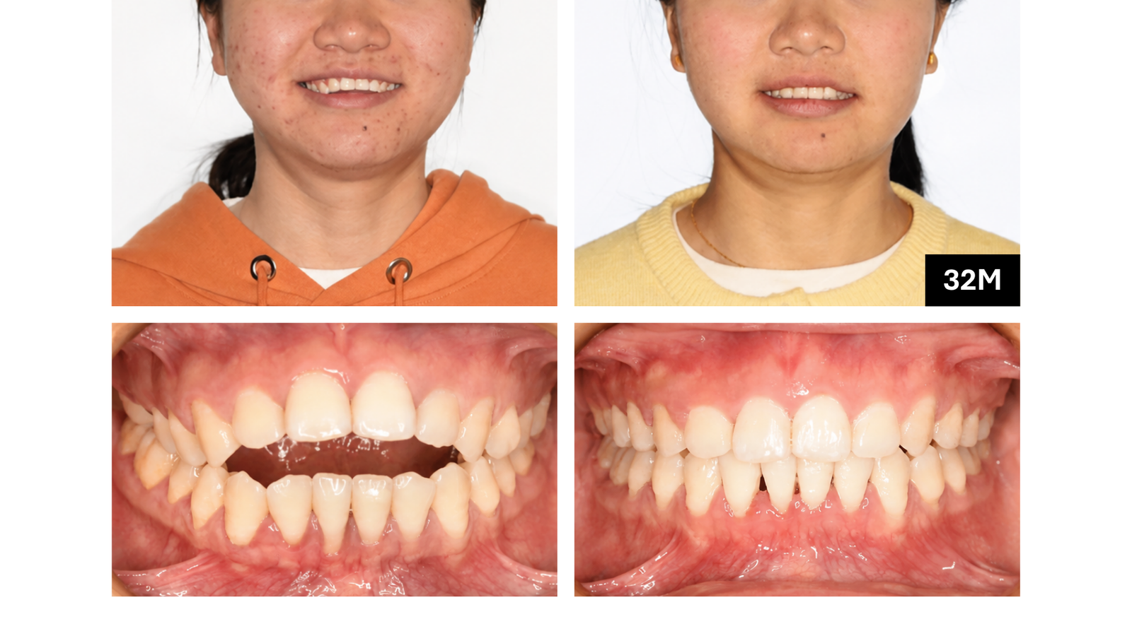
Figure 12. Frontal comparison before and after treatment, with the corresponding anterior occlusion
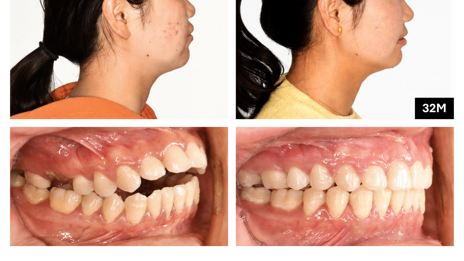
Figure 13. Profile comparison before and after treatment, with the corresponding buccal occlusion
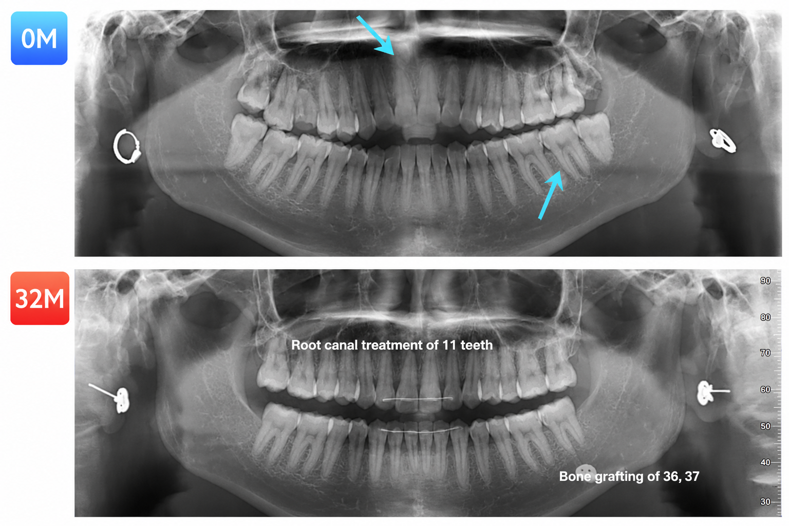
 

Figure 14. Panoramic radiographs at 0 and 32 months, annotated with the planned root canal therapy for 11 and resin restorations for 36 and 37
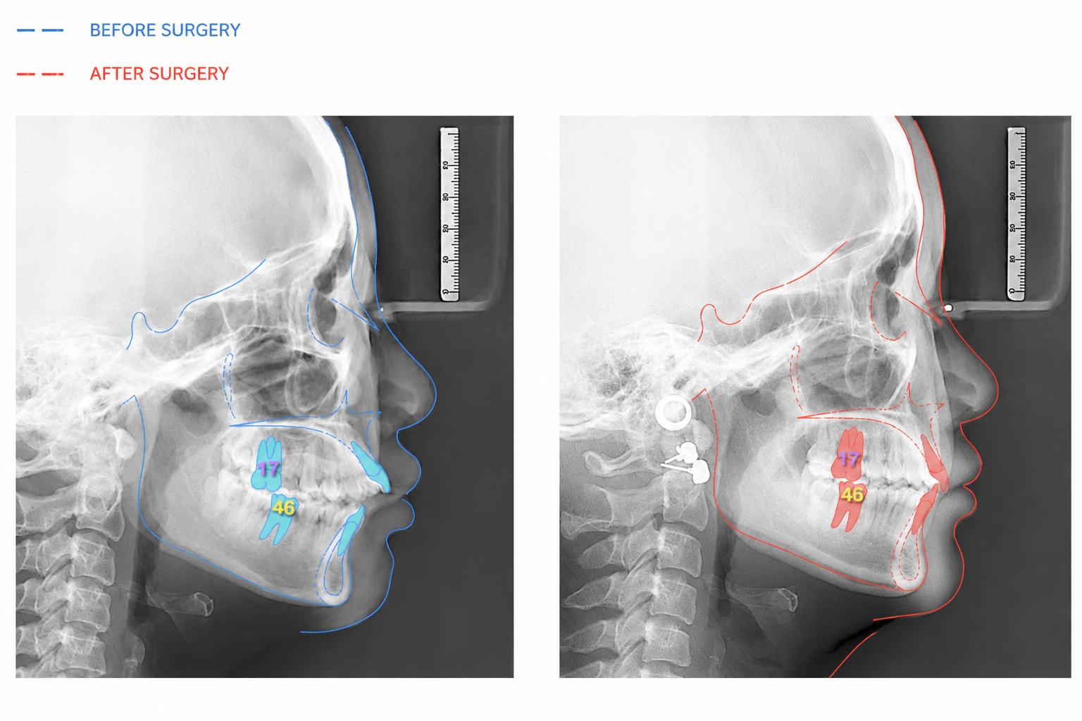
Figure 15. Cephalometric superimposition, blue indicates the pre-treatment tracing and red the post-treatment tracing
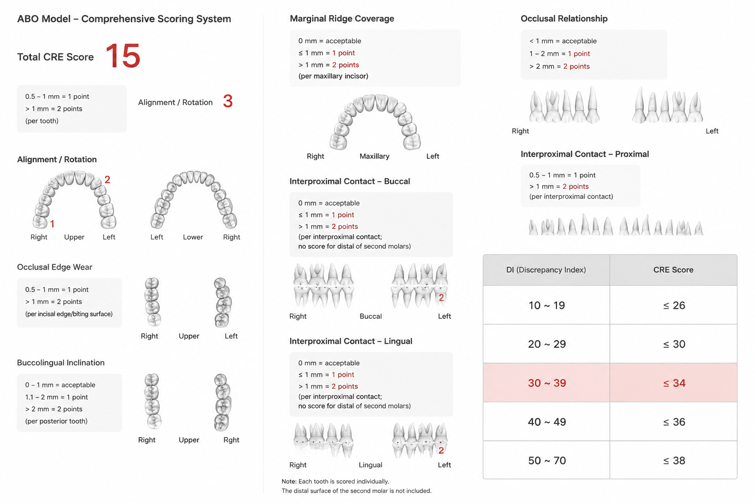
Figure 16. Completed ABO Cast-Radiograph Evaluation worksheet, total score 15 against a threshold of 34
 

Retention


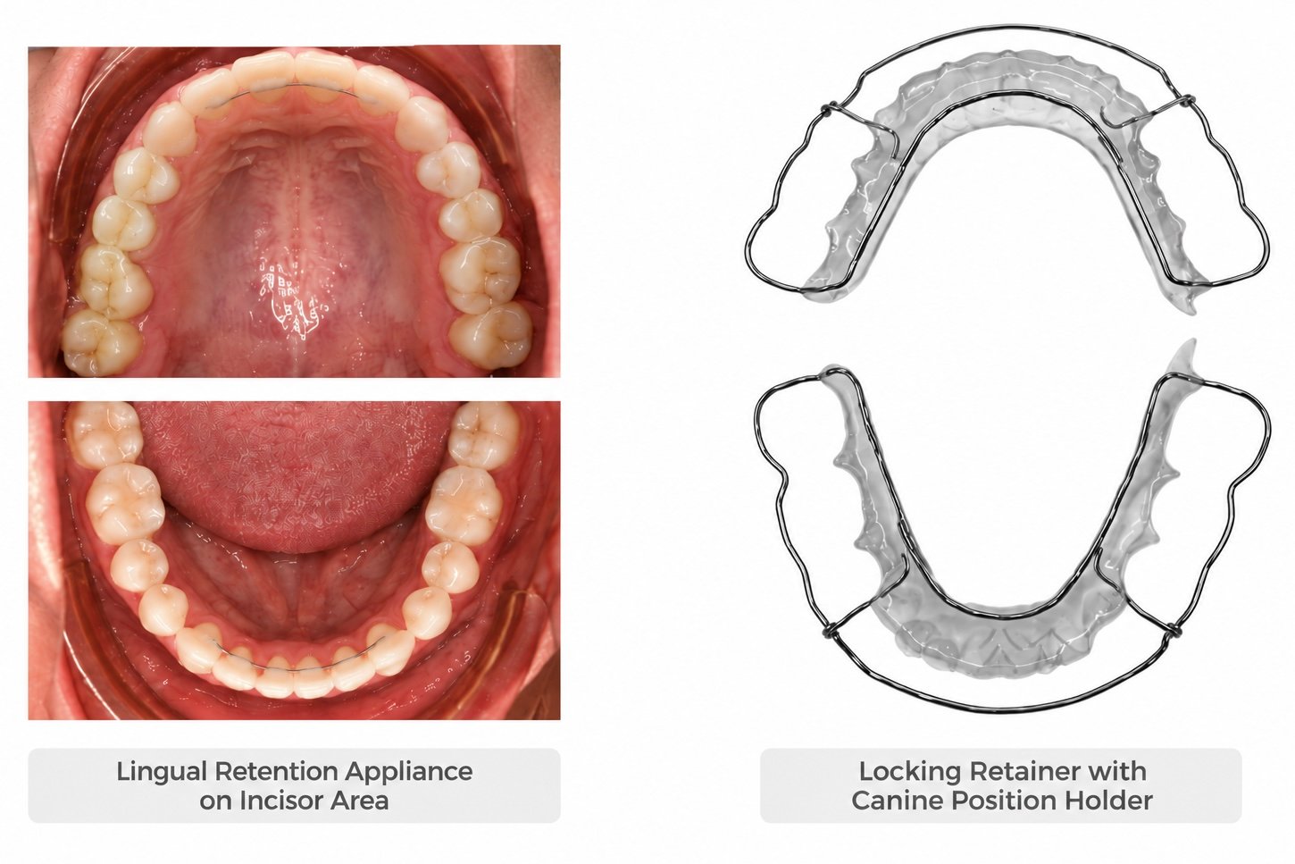
Open bite correction is vertically unstable by nature, so retention was planned specifically to hold the vertical result rather than simply to prevent rotational relapse. Two components were used together:

  • A bonded lingual retainer wire, to control the vertical position of the anterior teeth directly.
  • A Begg-type removable retainer, whose labial bow provides an additional vertical limiting effect on the incisors.

Conclusion

A severe anterior open bite in a normodivergent, skeletal Class I adult was closed completely without extracting a single premolar, and the large posterior space from a retained root was closed at the same time. Treatment took 32 months and finished with an ABO Cast-Radiograph Evaluation score of 15 against a threshold of 34.

The result depended on three things being done correctly and in the right order. First, the diagnosis: recognising a dental rather than a skeletal open bite is what made a non-surgical, non-extraction plan viable at all. Second, the mechanics: coordinating the arch forms removed the reverse curve, and relocating the occlusal fulcrum forward removed the posterior pivot that was holding the bite open — both of which reduced the open bite before skeletal anchorage was even engaged. Third, the anchorage: IZC and buccal shelf miniscrews delivered retraction, distalization and intrusion while holding the mandibular plane angle completely unchanged, which is what distinguishes a stable closure from a temporary one.

That the mandibular plane angle finished exactly where it started, after this much tooth movement, is the clearest evidence that the vertical dimension was controlled throughout rather than merely surviving treatment. Combined with a retention protocol designed specifically for vertical stability, it provides a sound basis for a lasting result.

Figure 23. Retention protocol. Left: bonded lingual retainer wire for vertical control. Right: Begg-type retainer with a labial bow providing vertical limitation.


Discussion: Distinguishing Skeletal from Dental Open Bite

The single most consequential decision in this case was made before any appliance was placed. The features that separate a skeletal from a dental open bite are summarised below.
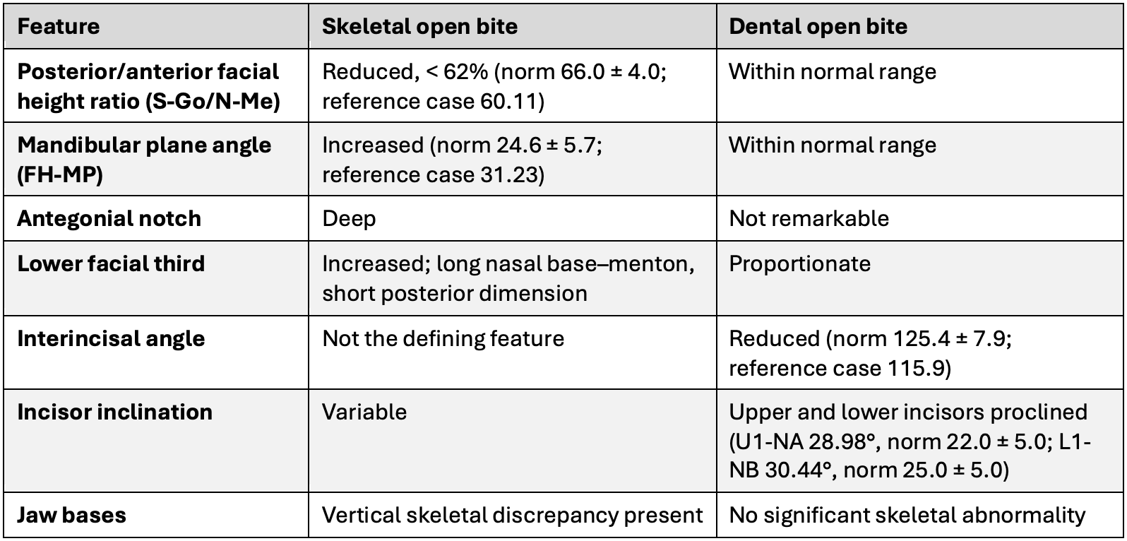
Table 7. Differential diagnosis of skeletal versus dental open bite. The numerical values in this table are drawn from the illustrative reference cases used in the original presentation, not from this patient.
 

Applying these criteria to the present patient: the mandibular plane angle was average (FMA 26°, within the normal range of 25 ± 6), the skeletal relationship was Class I (ANB 4°), and the jaw bases showed no significant abnormality. The abnormalities were confined to the incisors, which were proclined in both arches (U1-SN 111°, L1-MP 100°). This is the profile of a dental open bite, and it is what made a non-surgical, non-extraction approach realistic.

The clinical warning that follows from this comparison is worth stating plainly: a high mandibular plane angle combined with an open bite should always prompt caution, because that combination signals a skeletal component whose correction is far less predictable and considerably more prone to relapse.


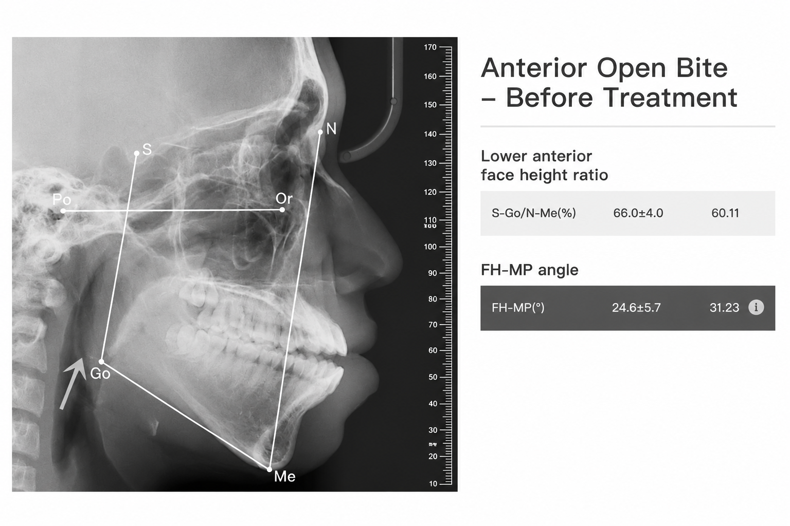
Figure 24. Reference case illustrating the features of a skeletal open bite: reduced posterior/anterior facial height ratio, increased mandibular plane angle, and a deep antegonial notch.
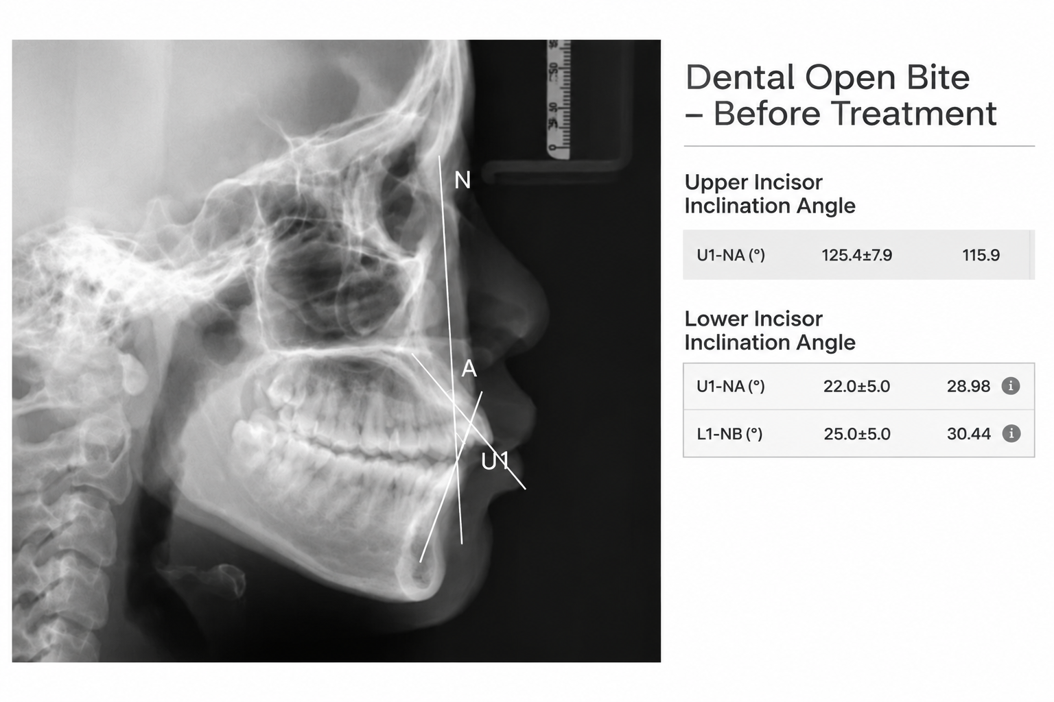
Figure 25. Reference case illustrating the features of a dental open bite: reduced interincisal angle and proclined upper and lower incisors, with no significant skeletal abnormality.

Cart (0 items)
Select the fields to be shown. Others will be hidden. Drag and drop to rearrange the order.
  • Image
  • SKU
  • Rating
  • Price
  • Stock
  • Availability
  • Add to cart
  • Description
  • Content
  • Weight
  • Dimensions
  • Additional information
Click outside to hide the comparison bar
Compare