

Introduction
Skeletal Class II malocclusion is among the most frequently encountered orthodontic problems in growing patients and is commonly associated with mandibular retrusion rather than maxillary protrusion. Left untreated, the discrepancy may lead to an excessive overjet, compromised occlusion, and an imbalanced facial profile. Early orthopedic intervention during the growth period provides an opportunity to stimulate mandibular development, reducing the severity of the skeletal discrepancy before comprehensive orthodontic treatment.
This case presents the two-phase treatment of a 12-year-old patient with skeletal Class II malocclusion. Growth modification using a Twin Block appliance was first employed to encourage mandibular advancement and improve the skeletal relationship. Once favorable jaw development had been achieved, comprehensive fixed appliance therapy supported by a digital indirect bonding workflow was carried out to refine tooth alignment, establish functional occlusion, and improve overall facial harmony.
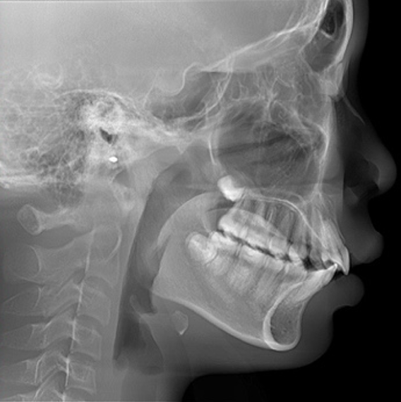
Figure 1. Pre-treatment lateral cephalogram
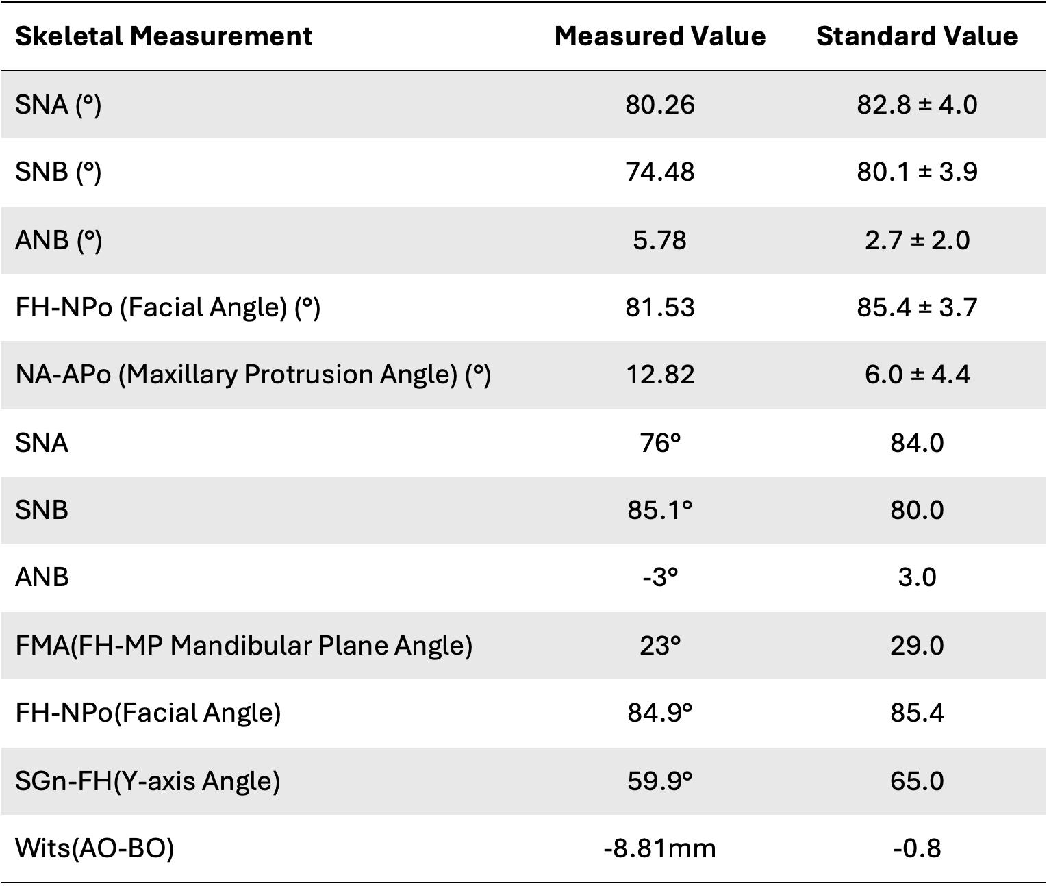
Table 1. Pre-treatment cephalometric analysis
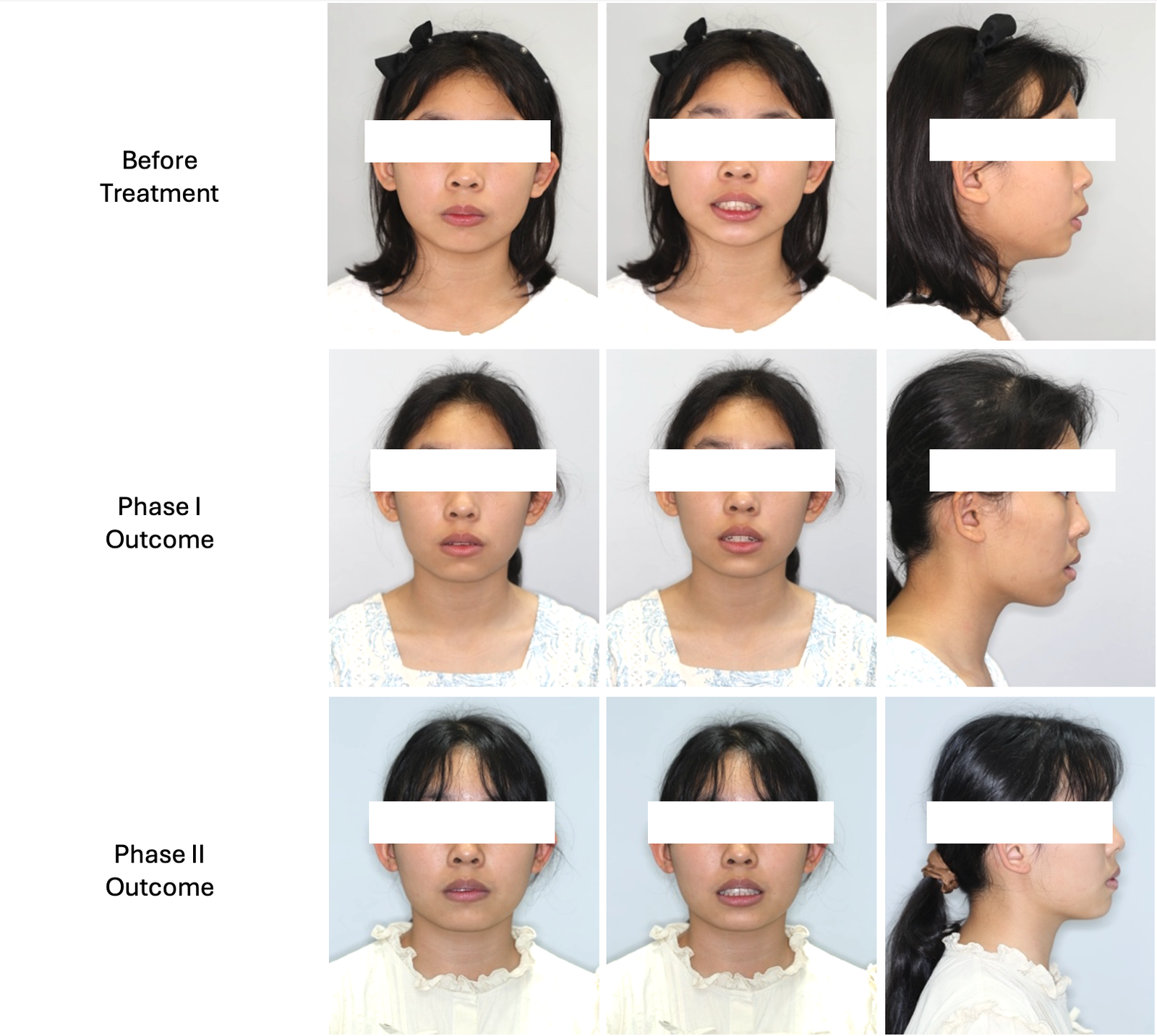
Figure 2. Facial appearance progression
Diagnosis
Initial clinical and cephalometric examination revealed a skeletal Class II malocclusion primarily associated with mandibular retrusion. The patient presented with a convex facial profile, retrusive chin, and favorable growth potential for orthopedic intervention.
Clinical Findings
- Mandibular Retrognathia (Mandibular Hypoplasia)
The mandible was positioned posteriorly relative to the cranial base, resulting in a retrusive lower jaw and convex facial profile. This finding is supported by a reduced SNB angle of 74.48° (normal ≈ 80°), indicating insufficient mandibular projection.
- Retrogenic Chin
The deficient mandibular growth resulted in inadequate chin prominence, contributing to reduced lower facial projection and overall facial imbalance.
- Low-angle Skeletal Pattern
The patient exhibited a relatively low mandibular plane angle (FMA: 25.3°), indicating a horizontal growth tendency and favorable potential for functional orthopedic treatment.
Cephalometric Findings
- Normal Maxillary Position
The SNA angle of 80.26° indicated that the maxilla was positioned within the normal range, suggesting that the skeletal discrepancy was not caused by maxillary protrusion.
- Skeletal Class II Relationship
The combination of a normal maxillary position and a retrusive mandible resulted in an increased ANB angle of 5.78°, confirming a skeletal Class II malocclusion primarily due to mandibular deficiency.
Objectives
Given the patient’s favorable growth potential, treatment was designed to address the skeletal discrepancy before focusing on dental correction. The primary objective was to promote mandibular advancement through functional orthopedic therapy, thereby improving the skeletal Class II relationship and facial profile. Following skeletal correction, comprehensive orthodontic treatment aimed to establish proper occlusion, achieve well-aligned dental arches, and create a stable, functional, and esthetic result.
Treatment Plans
To accomplish these objectives, treatment was divided into two sequential phases.
Phase I: Functional Orthopedic Treatment (14 Months)
The first phase focused on correcting the skeletal discrepancy by stimulating mandibular growth during the patient’s developmental period.
Treatment included:
- Twin Block functional appliance therapy to encourage mandibular advancement.
- Periodic appliance adjustments and monitoring of skeletal growth response.
- Continuous evaluation of occlusal development and facial profile improvement.
Following 14 months of functional appliance therapy, the patient demonstrated improved mandibular projection and a more favorable skeletal relationship, providing an ideal foundation for comprehensive orthodontic treatment.
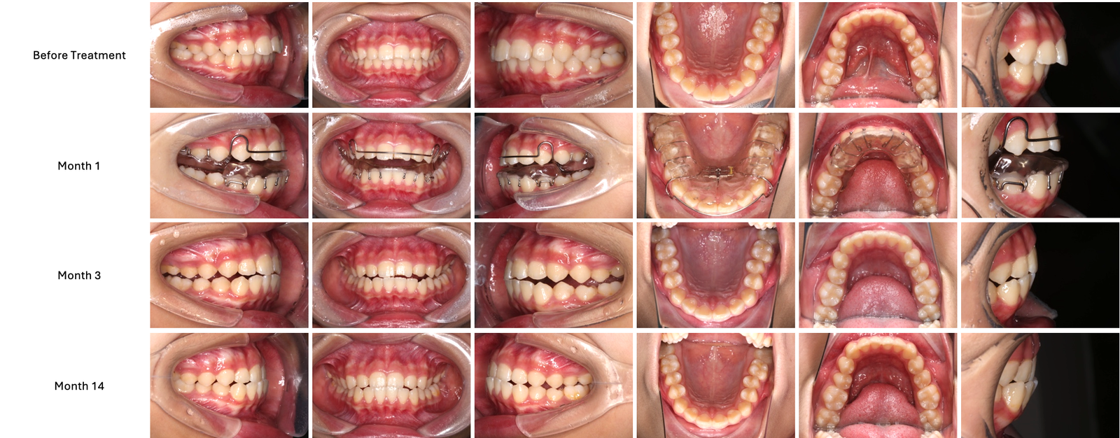
Figure 3. Case Progression (Phase I)
Phase II: Comprehensive Orthodontic Treatment (22 Months)
Following successful skeletal correction, comprehensive orthodontic treatment was initiated to establish functional occlusion and optimize dental alignment.
Treatment included:
- Extraction of teeth 38 and 48.
- Customized digital indirect bonding workflow for precise bracket placement.
- Alignment and leveling of the maxillary and mandibular dentitions.
- Placement of bilateral temporary anchorage devices (TADs) along the mandibular external oblique ridge to distalize the mandibular dentition.
- Mini-implant-supported Class II elastics for controlled retraction of the maxillary anterior teeth.
- Finishing and fine adjustment of the occlusal relationship.
- Adjunctive labial myofunctional training to improve functional stability.
The customized digital indirect bonding workflow enabled accurate bracket placement from the outset, improving treatment efficiency and allowing greater control over biomechanics throughout the comprehensive orthodontic phase.
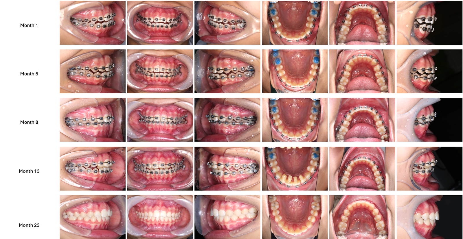
Treatment Outcomes
The staged treatment approach produced significant improvements in both skeletal and dental relationships. Functional orthopedic therapy successfully advanced the mandible, reducing the underlying skeletal Class II discrepancy and improving chin projection during the patient’s growth period. This created a more favorable skeletal foundation for the second phase of treatment.
Comprehensive orthodontic treatment subsequently achieved well-aligned dental arches, coordinated intercuspation, and a stable functional occlusion. Facial profile analysis also demonstrated improved lower facial balance and enhanced chin prominence. By combining early growth modification with precise digital orthodontic planning, treatment progressed efficiently and resulted in predictable functional and esthetic outcomes.
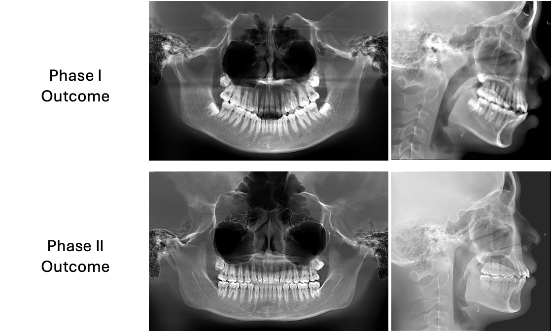
Figure 5. Post-treatment lateral cephalogram
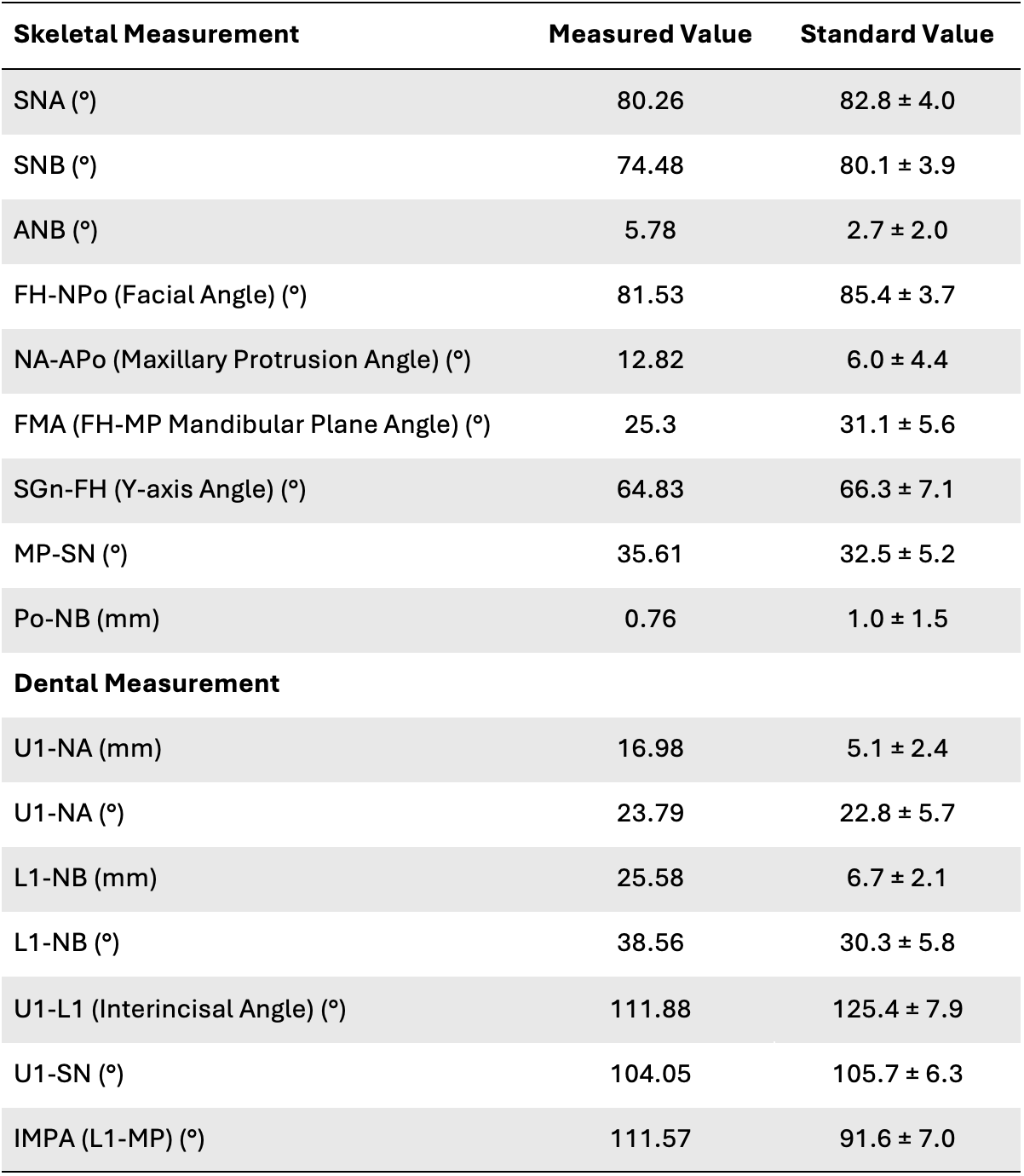
Table 2. Post-treatment cephalometric analysis
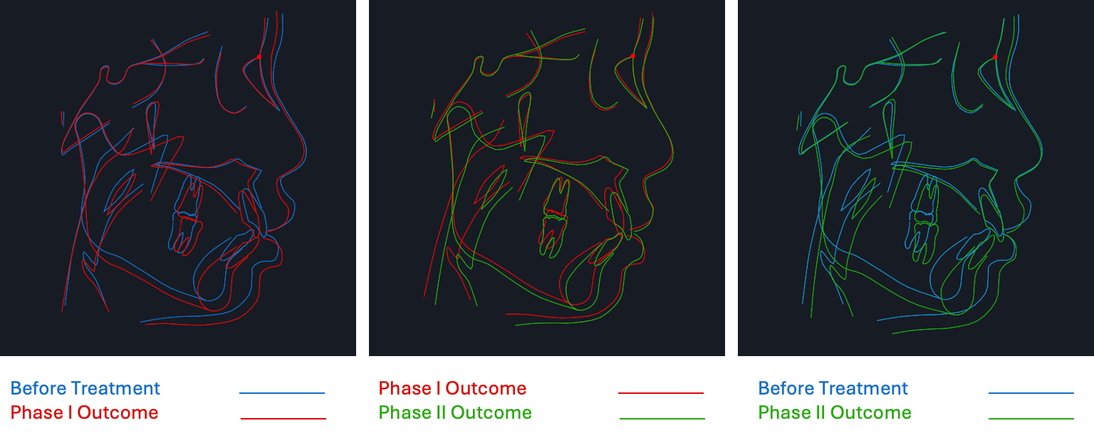
Figure 6. Cephalometric skeletal graph (pre- vs. post-treatment)
Conclusion
This case demonstrates how maximizing a patient’s remaining growth potential can fundamentally influence the outcome of skeletal Class II treatment. Through timely mandibular advancement, the underlying skeletal imbalance was improved before comprehensive orthodontic treatment, allowing the second phase to focus on achieving precise dental alignment, functional occlusion, and enhanced facial harmony rather than compensating for skeletal deficiency.
Supported by a digital indirect bonding workflow, treatment was carried out with greater precision and consistency, enabling efficient appliance placement and controlled biomechanics throughout fixed appliance therapy. By combining biologically appropriate growth modification with digitally guided treatment execution, clinicians can achieve predictable, individualized outcomes while optimizing both clinical efficiency and long-term treatment stability.