

A skeletal Class III malocclusion with a full anterior crossbite, an ANB of −3°, and a maxilla sitting well behind the cranial base is the kind of case that often ends in orthognathic surgery. This one did not. Through a structured two-phase protocol, the ANB was returned to near-normal, the anterior crossbite fully corrected, and a stable Class I occlusion established — the profile rebalanced by orthodontics alone.
This case study follows how that result was achieved. A 15-year-old patient with a deficient maxilla was treated across two coordinated phases — skeletal growth modification, then digitally guided fixed appliances using the ORTHGUIDE Indirect Bonding Guide — to reach a stable Class I occlusion and a non-surgical correction.
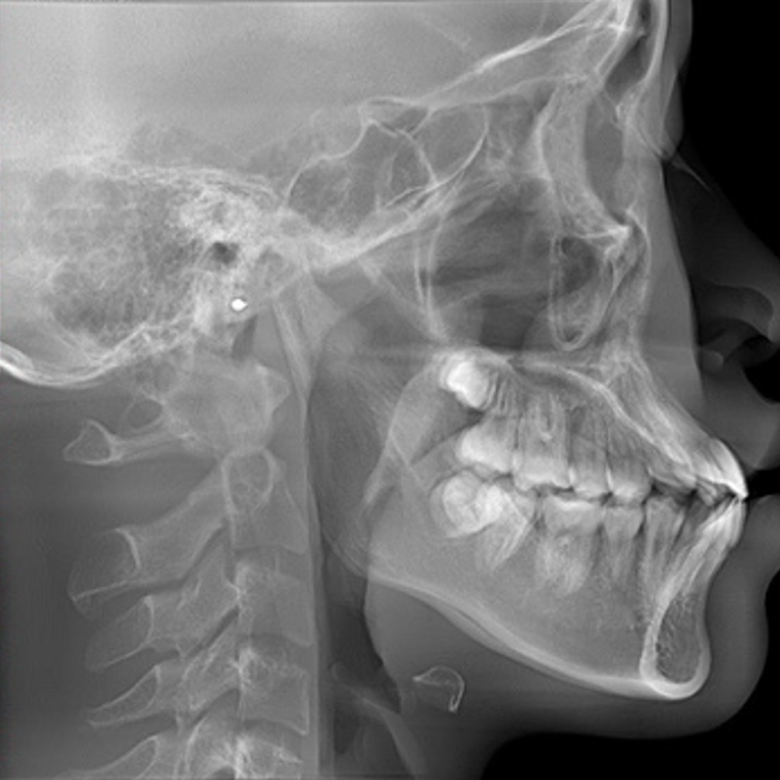
Figure 1. Pre-treatment lateral cephalogram (before treatment)
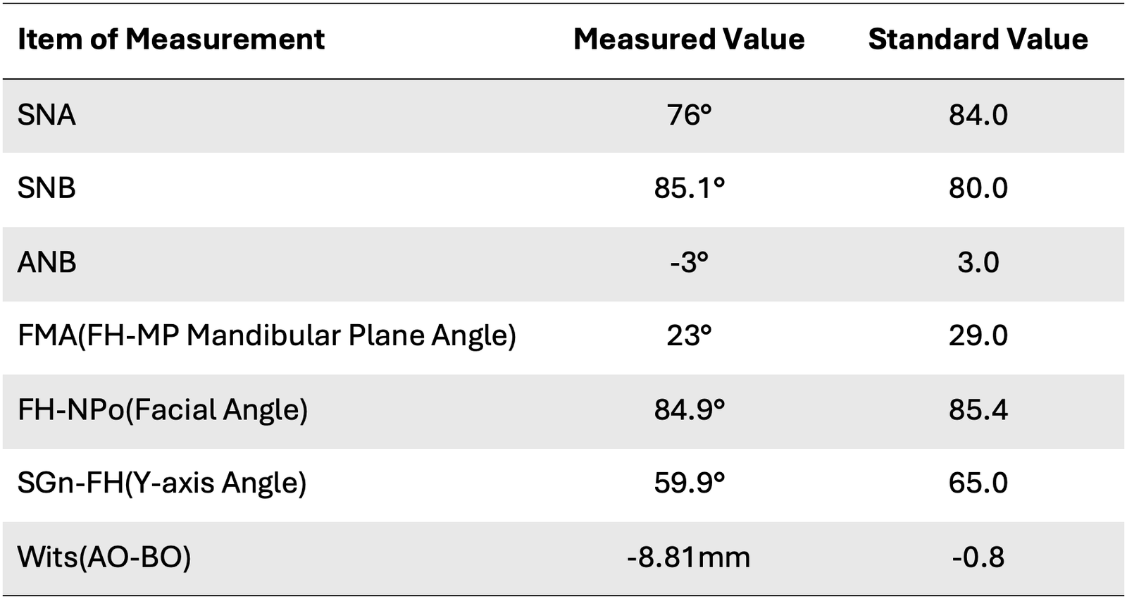
Table 1. Pre-treatment cephalometric analysis (before treatment)
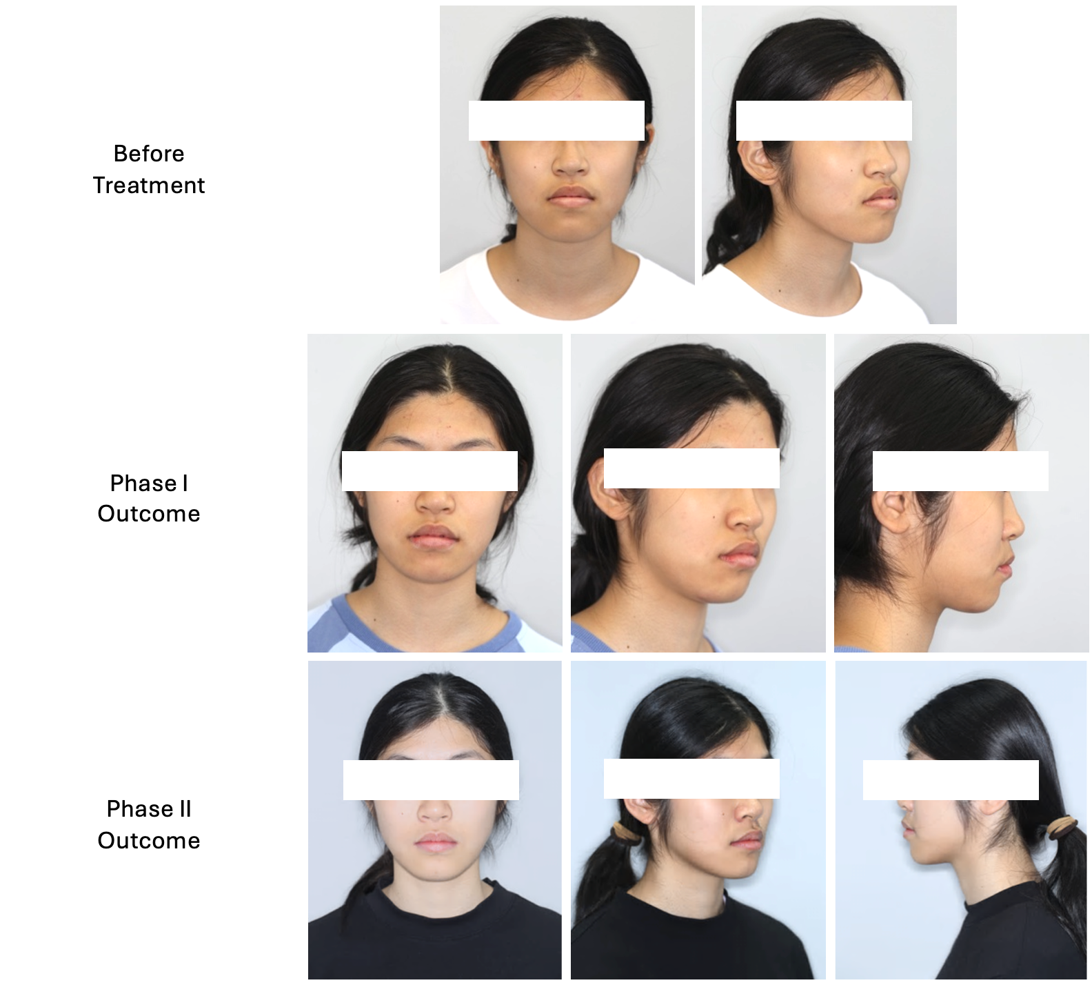
Figure 2. Facial appearance progression
Initial clinical and cephalometric analysis revealed a severe skeletal Class III malocclusion characterized by:
- Anterior Crossbite
The maxillary anterior teeth occlude behind the mandibular anterior teeth due to the underlying skeletal Class III discrepancy, resulting in compromised occlusion and facial esthetics.
- Maxillary Transverse Deficiency
The maxillary arch is narrower than the mandibular arch, contributing to inadequate transverse width, posterior crossbite, and insufficient space for ideal dental alignment.
- Maxillary Hypoplasia
Underdevelopment of the maxilla results in a retrusive upper jaw. This is supported by a reduced SNA angle of 76.0° (normal ≈ 82–84°), indicating deficient maxillary projection.
- Mandibular Hyperplasia
Excessive mandibular growth causes a protrusive lower jaw and contributes to the skeletal Class III relationship. This is reflected by an increased SNB angle of 85.1° (normal ≈ 80°).
The combination of maxillary retrusion and mandibular prognathism is confirmed by an ANB angle of −3.0° and a Wits appraisal of −8.81 mm, indicating a significant skeletal Class III discrepancy. A relatively low mandibular plane angle (FMA: 23.0°) suggests that the patient also exhibited a horizontal growth pattern.
Objectives
The treatment aimed to facilitate forward growth of the maxilla to reduce the skeletal Class III discrepancy while taking advantage of the patient’s remaining growth potential. A secondary objective was to establish proper occlusion by correcting the anterior crossbite, improving the skeletal and dental relationships, and achieving stable intercuspation with functional and esthetic outcomes.
Treatment Plans
Treatment was delivered in two coordinated phases spanning roughly 28 months of active care.
Phase I: Orthopedic Correction (12 Months)
The first phase focused on early skeletal correction through growth modification.
Treatment included:
- Maxillary skeletal expansion (MSE) to address transverse deficiency
- Maxillary protraction to stimulate forward maxillary development
- Continuous monitoring of skeletal response and occlusal improvement
This phase successfully improved the patient’s maxillary position while reducing the severity of the skeletal Class III relationship, creating a more favorable foundation for comprehensive orthodontic treatment.
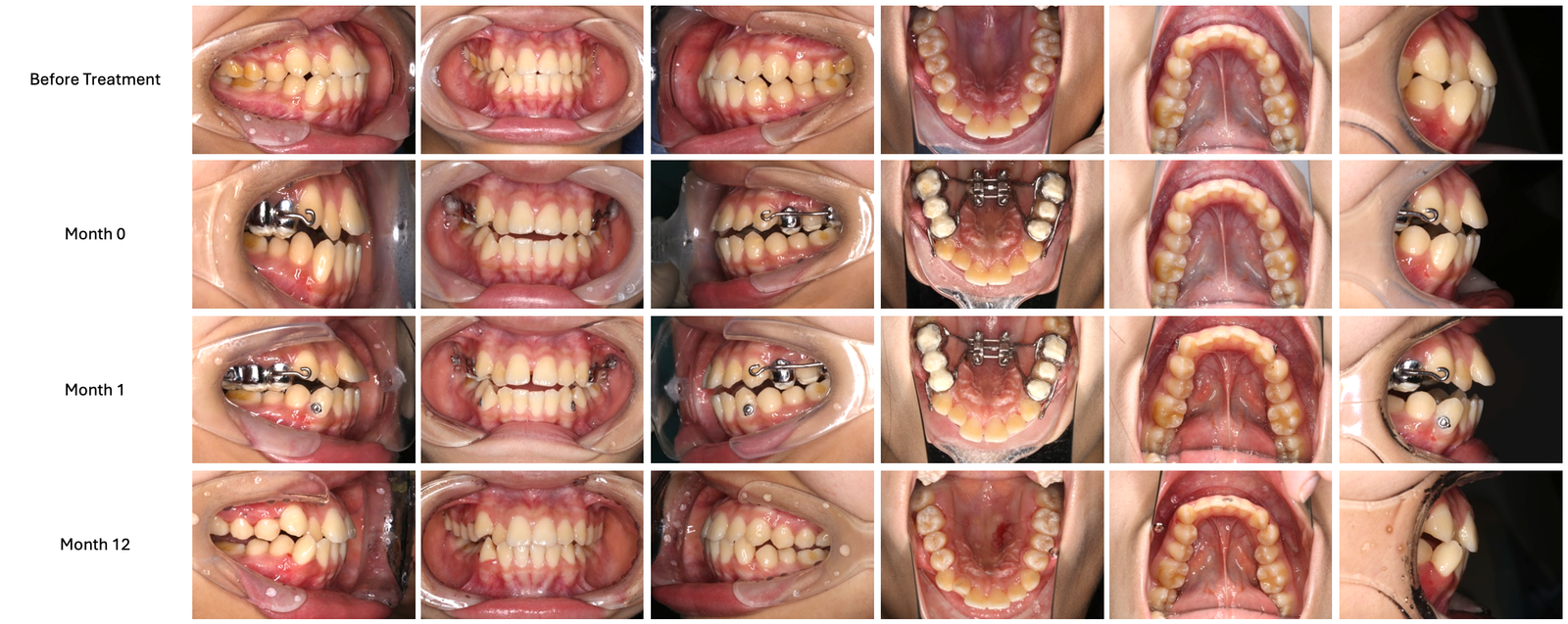
Figure 3. Case Progression (Phase I)
Phase II: Comprehensive Orthodontic Treatment (17 Months)
Following orthopedic correction, comprehensive orthodontic treatment was initiated to refine occlusion and establish long-term stability.
Treatment included:
- Extraction of teeth 17, 27, 38, and 48
- Customized digital positioning guide using the OrthGuide Indirect Digital Bonding (IDB) System
- Alignment and leveling of both arches
- Bite turbos on the maxillary anterior teeth to eliminate localized anterior crossbite
- Placement of bilateral mandibular temporary anchorage devices (TADs)
- Mandibular dentition distalization using TAD-supported mechanics combined with Class II elastics
- Fine occlusal detailing and finishing
- Adjunctive labial myofunctional therapy
The customized digital positioning guide enabled accurate appliance placement and enhanced control throughout treatment, supporting efficient biomechanics and predictable tooth movement.
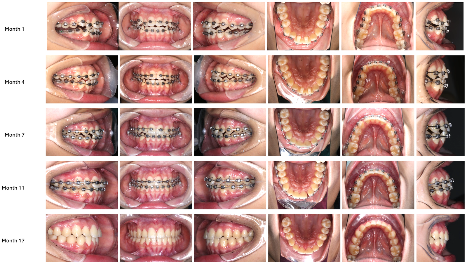
Figure 4. Case Progression (Phase II)
Treatment Outcomes
Following completion of both treatment phases, the patient demonstrated substantial improvements in skeletal, dental, and functional relationships.
Key outcomes included:
- Significant improvement in skeletal Class III discrepancy
- Correction of anterior crossbite
- Improved maxillary position and facial profile
- Establishment of functional occlusion with favorable intercuspation
- Enhanced dental alignment and arch coordination
- Stable treatment results with improved esthetics and function
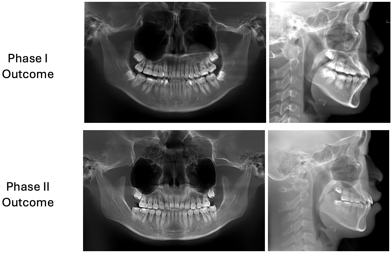
Figure 5. Pre-treatment lateral cephalogram (after treatment)
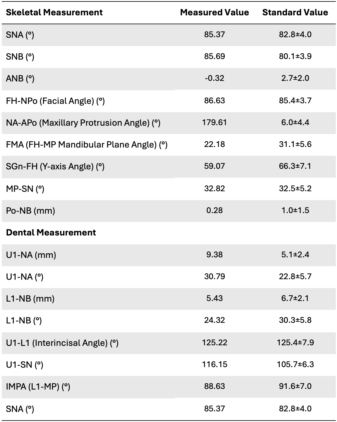
Table 2. Pre-treatment cephalometric analysis (after treatment)
By the end of Phase II, cephalometric analysis confirmed substantial skeletal improvement, with the ANB angle improving from −3° before treatment to −0.32° after treatment, reflecting successful orthopedic correction and orthodontic finishing.
Conclusion
Successful management of skeletal Class III malocclusion relies on accurate diagnosis, timely orthopedic intervention, and meticulous biomechanical control. In this case, a staged treatment approach effectively corrected the underlying skeletal discrepancy, established functional occlusion, and improved overall facial harmony.
By integrating a digital workflow and a customized Indirect Bonding System into treatment, greater precision was achieved during appliance placement and treatment execution, allowing for efficient biomechanics and predictable clinical outcomes. As digital technologies continue to evolve, they offer valuable support in enhancing treatment accuracy, consistency, and overall clinical efficiency in complex orthodontic cases.
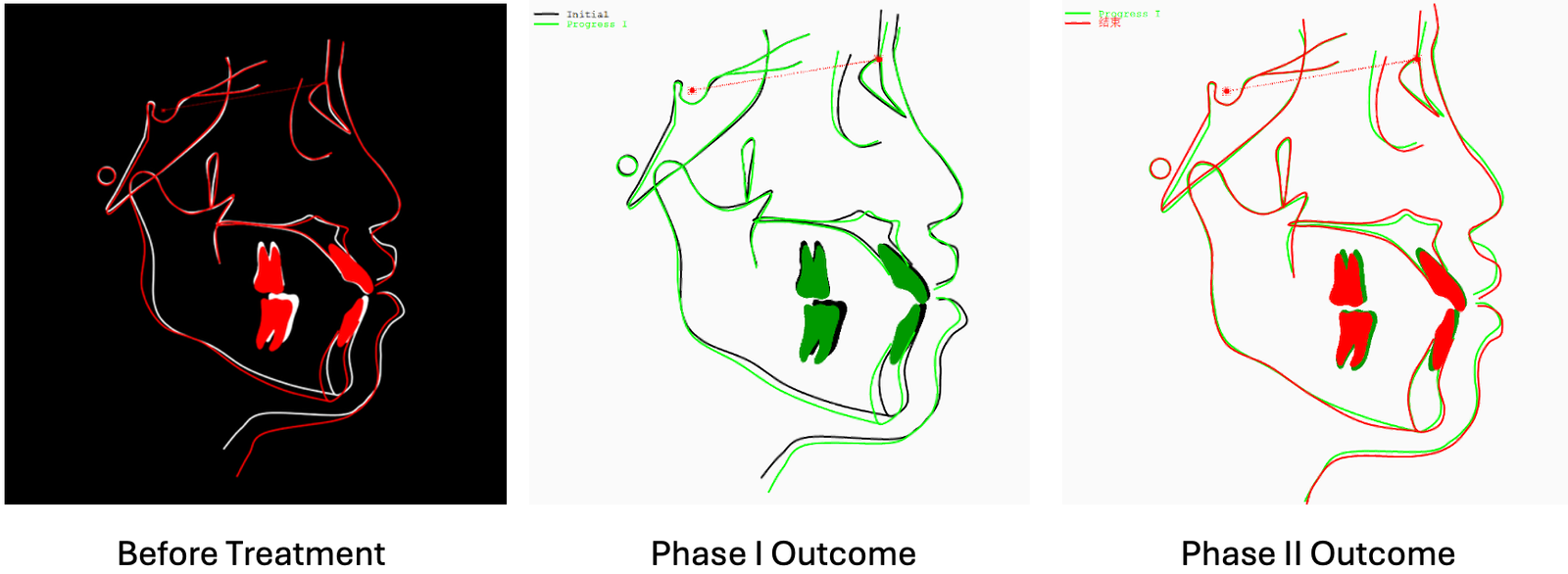
Figure 6. Cephalometric skeletal graph (before vs. after treatment)